Understanding the fundamentals of diagnostic imaging is crucial for medical students and professionals aiming to excel in clinical practice. A solid grasp of this subject enables effective patient care and accurate decision-making in various medical scenarios. With numerous resources available, it can be challenging to navigate the vast amount of material efficiently. This guide offers valuable insights into how to approach the preparation process with confidence and focus.
The key to success lies in identifying the core concepts and honing the skills necessary to interpret complex medical visuals. By familiarizing yourself with common techniques, terminology, and approaches, you will build a foundation that enhances both your theoretical knowledge and practical capabilities. Engaging with practice materials and reviewing frequently tested content can further solidify your understanding.
In this article, we will explore essential strategies to help you master the subject and tackle related challenges with ease. Whether you’re preparing for assessments or refining your clinical judgment, these insights will serve as a helpful guide to achieving your goals.
Aquifer Radiology Exam Answers
Preparing for assessments related to diagnostic imaging requires both theoretical knowledge and practical understanding of the visual techniques used in clinical practice. Mastery of these concepts helps you accurately analyze and interpret various medical visuals, which is essential for providing optimal patient care. To succeed, it’s crucial to familiarize yourself with the format of questions typically encountered and to approach each case scenario methodically.
Focusing on core principles such as image interpretation, common conditions, and the key markers used in diagnostics can significantly boost your confidence. A thorough review of frequently tested topics and clinical scenarios will allow you to anticipate the types of questions that may arise, improving your chances of success. Building a structured study routine that integrates practical application and theoretical knowledge is an effective approach to mastering the material.
Utilizing a range of resources, from textbooks to practice exercises, can deepen your understanding and reinforce important concepts. Engaging with these materials not only sharpens your skills but also helps you recognize patterns in diagnostic imaging. With consistent effort, you’ll be well-equipped to excel in any related assessment, ensuring you’re ready to apply your knowledge in real-world situations.
Understanding Radiology Exam Format
Familiarity with the structure of clinical assessments focused on imaging is essential for success. These tests are designed to evaluate not only your theoretical knowledge but also your ability to apply concepts in real-world scenarios. Understanding the format of these evaluations will help you approach each question methodically, increasing your chances of achieving a high score.
Typically, these assessments include a mix of question types, such as:
- Multiple-choice questions (MCQs) that test your knowledge of diagnostic techniques and terminology.
- Case-based questions where you must analyze images and provide a diagnosis or treatment plan.
- Short-answer questions focusing on key concepts or specific conditions.
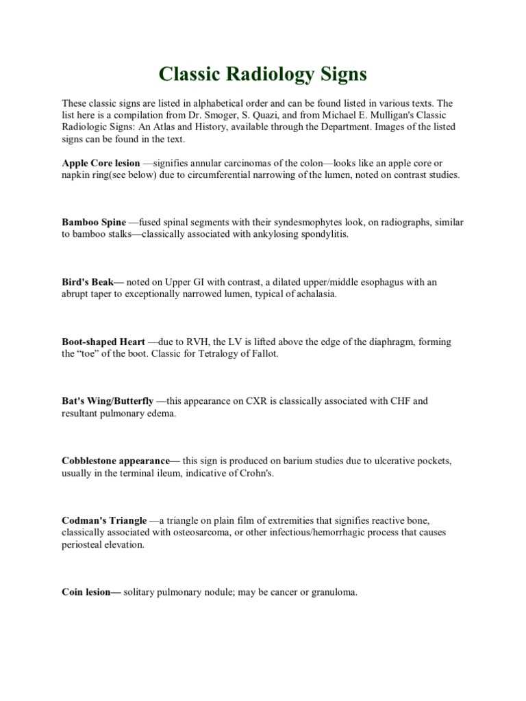
It’s important to recognize that some questions will require not just recalling facts, but also interpreting clinical visuals. Being able to analyze images, identify anomalies, and link them to relevant conditions is a skill that’s highly valued in these tests.
In addition, managing time efficiently is critical. Each question or section of the test may vary in difficulty and length, so understanding how to pace yourself throughout the assessment is key to ensuring you have enough time to address all areas.
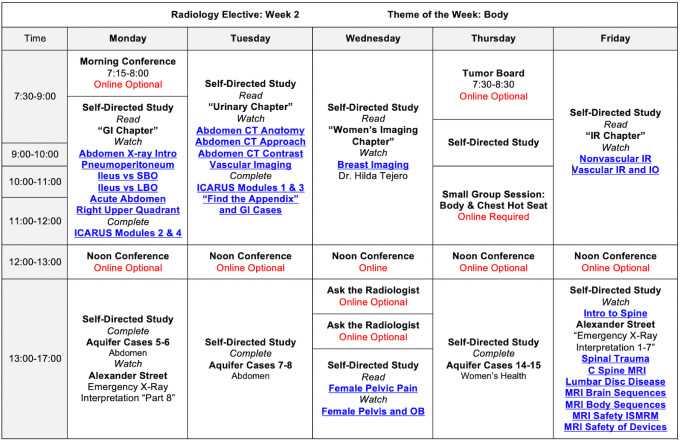
Preparation should focus on both theory and practice. Reviewing common conditions, imaging modalities, and diagnostic markers can help you feel more confident when faced with these types of questions. Engaging in mock assessments or practice tests is a great way to familiarize yourself with the test’s flow and time constraints.
Key Concepts in Diagnostic Imaging
Grasping the essential principles of diagnostic imaging is fundamental for interpreting medical visuals accurately. These concepts form the backbone of many clinical assessments and are critical for making informed decisions in patient care. By understanding key topics, you can develop a deeper understanding of how imaging works and how to interpret various conditions that may be present in a patient’s scans.
Fundamental Imaging Techniques
There are several primary techniques used in clinical imaging, each serving a different purpose in diagnosis. Familiarity with these methods is crucial for recognizing and understanding the images you encounter:
- X-rays: Commonly used to detect bone fractures, infections, or abnormalities in lung tissue.
- CT scans: Provide detailed cross-sectional images that help diagnose complex conditions in organs, tissues, and bones.
- Ultrasound: Used for evaluating soft tissues, blood flow, and organ functions in a non-invasive way.
- MRI: Provides detailed images of soft tissues, helping in the diagnosis of neurological, muscular, and joint issues.
Common Medical Conditions to Identify
Familiarity with common conditions that are often identified through imaging is essential. Some of the most frequently diagnosed issues include:
- Fractures: Broken bones that often require immediate intervention and can be clearly seen on X-rays.
- Tumors: Abnormal growths that may appear in different imaging techniques, requiring further investigation to assess malignancy.
- Infections: Conditions such as pneumonia or abscesses that can be detected through various imaging methods, especially X-rays and CT scans.
- Arterial blockages: Identified through techniques like ultrasound or CT angiography to evaluate blood flow.
By mastering these foundational techniques and recognizing common conditions, you’ll be well-prepared for interpreting clinical visuals in assessments and real-world practice.
Common Mistakes During Radiology Exams
During assessments that involve the interpretation of medical images, several common pitfalls can hinder success. These errors often arise from misunderstandings of key concepts or misapplication of techniques, and they can significantly impact your ability to accurately diagnose or analyze the given case. Recognizing these mistakes is essential to avoid them and to refine your approach to clinical assessments.
Below is a table highlighting some of the most frequent errors made during these evaluations and tips on how to avoid them:
| Common Mistake | Description | How to Avoid It |
|---|---|---|
| Misinterpreting Image Orientation | Failing to correctly assess the positioning of the image can lead to misdiagnosis, especially with X-rays and CT scans. | Always check the markers indicating the image orientation before proceeding with the analysis. |
| Overlooking Subtle Signs | Small but critical details can often be missed, such as early-stage abnormalities or small fractures. | Take time to carefully examine each part of the image, looking for even the faintest signs of issues. |
| Relying Too Heavily on One Image | Making a diagnosis based solely on a single image or view can lead to incomplete or incorrect conclusions. | Review all available images and ensure a comprehensive assessment from multiple angles. |
| Ignoring Patient History | Without considering the patient’s clinical background, it’s easy to misinterpret certain findings in the images. | Always correlate the images with the patient’s history and symptoms for a more accurate diagnosis. |
| Underestimating the Importance of Contrast | Not fully understanding how contrast agents affect image clarity can lead to misreading important details. | Ensure you understand the role of contrast agents and their effects on the imaging process. |
By being aware of these common errors and taking proactive steps to avoid them, you can improve your performance and accuracy during assessments involving medical imaging.
Effective Study Tips for Success
To excel in assessments that require the interpretation of medical images, a structured and focused approach to studying is essential. Effective preparation not only strengthens your understanding of key concepts but also builds the confidence necessary to perform well in challenging scenarios. By utilizing a variety of strategies, you can optimize your study sessions and improve your chances of success.
The following table outlines some proven study tips that can help you maximize your learning and perform at your best:
| Study Tip | Description | How to Implement |
|---|---|---|
| Active Recall | Engaging with the material by testing yourself frequently to reinforce memory retention. | After reviewing a topic, close your materials and try to recall key points without looking at your notes. |
| Practice with Real Images | Working with actual medical visuals helps bridge the gap between theory and practical application. | Use online resources or textbooks with case studies to practice identifying conditions from images. |
| Spaced Repetition | Reviewing information at spaced intervals to improve long-term retention. | Use flashcards or apps designed for spaced repetition to revisit concepts at increasing intervals. |
| Group Study | Collaborating with peers can provide diverse insights and improve your understanding of complex topics. | Join a study group where you can discuss case studies and quiz each other on different conditions. |
| Time Management | Allocating specific time blocks for focused study sessions to avoid burnout and maximize productivity. | Set a timer for focused study sessions (e.g., Pomodoro technique) and take breaks to refresh your mind. |
By incorporating these strategies into your study routine, you can enhance your preparation and approach the assessment with greater confidence and competence.
How to Approach Case Scenarios
When presented with clinical case scenarios, it’s essential to adopt a systematic approach to analyze the situation effectively. These cases often require not just recalling facts, but also the ability to apply knowledge to diagnose conditions or propose treatment plans based on available data. Developing a structured method to assess each case can help ensure a thorough and accurate response.
Steps for Analyzing Case Scenarios
Breaking down case scenarios into manageable steps allows you to focus on relevant details while avoiding common errors. Here are key stages to follow when tackling these scenarios:
| Step | Description | Action |
|---|---|---|
| 1. Read the Case Carefully | Ensure you understand all aspects of the case, including patient history and symptoms. | Highlight important details such as age, medical history, and key complaints. |
| 2. Identify Key Clinical Signs | Look for visual clues or physical symptoms that indicate potential conditions. | Pay attention to specific patterns or abnormalities described in the case. |
| 3. Narrow Down Potential Diagnoses | Using your knowledge, list possible diagnoses based on the evidence presented. | Eliminate unlikely conditions by ruling out symptoms that don’t align with the case. |
| 4. Formulate a Diagnostic Plan | Determine what additional information or tests are needed to confirm your diagnosis. | Recommend the appropriate tests or imaging techniques to narrow down the diagnosis. |
| 5. Consider Treatment Options | Once a diagnosis is made, think through the appropriate treatment strategies. | Weigh the benefits of different approaches and consider patient preferences. |
Tips for Success in Case Scenarios
In addition to following a structured approach, here are a few tips to enhance your performance:
- Stay calm and avoid rushing through the case.
- Consider the patient’s overall health and context in your decision-making.
- Review relevant anatomy and physiology to better understand the implications of the case.
- Look for clues in the wording of the case that may suggest specific conditions or complications.
By following these guidelines, you can approach case scenarios with greater confidence and precision, ensuring that your analysis is both thorough and accurate.
Essential Medical Imaging Terms to Know
Understanding the language of medical imaging is crucial for interpreting diagnostic images accurately. Various terms are commonly used in the field to describe techniques, findings, and conditions that can be identified through imaging. Familiarity with these terms enhances your ability to communicate effectively and make informed decisions during assessments.
Below are some essential terms that you should be familiar with when working with imaging data:
- Contrast Agents: Substances used to enhance the visibility of certain tissues or structures in imaging scans. These agents help highlight abnormalities that might otherwise be difficult to detect.
- Modality: Refers to the type of imaging technique used, such as X-ray, CT scan, MRI, or ultrasound.
- Resolution: The ability of an imaging system to distinguish between small structures or details. Higher resolution typically provides clearer, more detailed images.
- Artifact: Distortions or errors in an image caused by technical issues or patient movement during the imaging process.
- Localization: Refers to determining the exact location of a suspected abnormality or condition within the body.
- Radiopaque: Describes substances or structures that do not allow X-rays or other imaging techniques to pass through, appearing white or light on the image.
- Radiolucent: Refers to areas or materials that allow imaging waves to pass through, appearing darker on the image.
- Lesion: Any abnormal tissue or growth, typically detected through imaging, that may be benign or malignant.
- Incidence: The frequency or occurrence of a particular finding or condition in a population, often used to describe the likelihood of detecting certain diseases through imaging.
- Cross-sectional Imaging: Techniques, such as CT and MRI, that create detailed, slice-like images of the body from different angles to allow for a 3D view.
By mastering these terms and concepts, you’ll be better prepared to navigate medical imaging challenges and make informed clinical decisions.
What to Expect in Assessments
When preparing for assessments in medical imaging and diagnostic interpretation, it’s important to understand the structure and expectations of these evaluations. These assessments are designed to test your ability to analyze medical images, recognize abnormalities, and make informed decisions based on the data presented. You’ll be challenged to apply theoretical knowledge to practical scenarios, all while ensuring that your decision-making process is both accurate and efficient.
During these evaluations, you can expect to encounter a variety of case studies that require you to interpret imaging results, diagnose conditions, and suggest appropriate management or treatment strategies. The cases will vary in complexity, requiring you to demonstrate both foundational knowledge and advanced diagnostic skills. Preparation for these assessments involves not just memorizing facts, but also honing your critical thinking and problem-solving abilities.
Typically, the assessment will include:
- Multiple case-based scenarios with accompanying medical images.
- Questions designed to test your diagnostic reasoning and ability to identify abnormalities.
- Practical components that challenge your understanding of the relationships between clinical findings and imaging results.
- Timed sessions to assess your ability to think quickly and accurately under pressure.
In addition, you may encounter interactive elements such as quizzes or feedback systems that provide immediate responses to your decisions, helping you learn as you progress through the material. Overall, these assessments are designed to simulate real-world clinical situations and evaluate how well you can integrate your knowledge into practice.
Analyzing Diagnostic Imaging Questions
When presented with questions related to medical images, it’s essential to approach them with a structured and analytical mindset. The goal is to extract key information from the provided images and clinical context to make accurate diagnoses or decisions. A systematic method of analysis helps in interpreting the images effectively, while also ensuring that no crucial details are overlooked.
To effectively analyze these types of questions, it’s important to break them down into manageable components. Here are key steps to consider when evaluating diagnostic imaging-related questions:
- Review the Clinical History: Always begin by considering the patient’s clinical history and symptoms. This context can provide clues that will guide your interpretation of the images.
- Identify the Key Image Features: Focus on identifying the most significant features in the image, such as areas of abnormality, unusual patterns, or unexpected findings.
- Formulate Differential Diagnoses: Based on the clinical history and image findings, come up with a list of potential conditions that could explain the observed abnormalities. Narrow down the possibilities based on further analysis.
- Apply Diagnostic Criteria: Use your knowledge of common conditions and diagnostic criteria to evaluate the most likely causes of the observed abnormalities. This step often involves applying diagnostic guidelines or algorithms.
- Consider Additional Testing: If the diagnosis is unclear, think about what further tests or imaging could help confirm or rule out certain conditions. Imaging often requires complementary tests to reach a definitive conclusion.
By following this approach, you ensure that each question is analyzed thoroughly and that your conclusions are based on both the image itself and the relevant clinical information. In doing so, you can arrive at more accurate interpretations and better-informed decisions.
Time Management for Imaging Assessments

Efficient time management is crucial when preparing for and taking imaging-related assessments. Balancing accuracy and speed can be challenging, but with proper strategies, you can improve your ability to work under time constraints. The key is to prioritize tasks, avoid unnecessary delays, and remain focused throughout the assessment process.
To manage your time effectively, consider the following approaches:
- Prioritize Key Tasks: Identify the most critical aspects of the assessment, such as the clinical history and image analysis, and focus your time and attention on them first.
- Allocate Time for Each Section: Divide the total time available into sections based on the number of tasks or questions. Stick to these time limits to avoid spending too much time on any one area.
- Familiarize Yourself with Question Formats: Understanding the typical structure of questions can help you anticipate the time required to answer them. This allows for faster decision-making during the assessment.
- Practice Under Time Pressure: Regularly practice with timed mock scenarios to get used to answering questions efficiently within a set period. This will help build confidence and improve your speed.
- Stay Calm and Focused: Remaining calm under time pressure is essential for making accurate decisions. Take deep breaths and focus on the task at hand to avoid rushing and making avoidable mistakes.
By implementing these strategies, you can enhance your ability to manage time effectively during imaging assessments, allowing you to maximize your performance and minimize stress.
Preparing for Difficult Medical Imaging Topics
Certain subjects within medical imaging can present significant challenges due to their complexity or the depth of knowledge required to fully understand them. These topics often involve intricate details, subtle distinctions in imaging findings, or advanced diagnostic techniques. Successfully preparing for these areas requires a focused approach, a willingness to invest time, and the use of varied learning strategies.
To tackle difficult topics, consider the following preparation strategies:
- Break Down Complex Concepts: Start by breaking down difficult topics into smaller, more manageable sections. Focus on understanding one aspect before moving on to the next. This will help build a solid foundation.
- Use Visual Aids: Medical imaging is inherently visual. Using annotated images, diagrams, or video tutorials can help solidify your understanding and highlight key features you need to focus on.
- Study in Small Chunks: Rather than overwhelming yourself with long study sessions, break your study time into smaller, focused intervals. This technique, known as “chunking,” can improve retention and reduce burnout.
- Practice with Case Scenarios: Applying your knowledge to real-world scenarios is one of the best ways to solidify your understanding. Practice with sample cases to test your diagnostic abilities and refine your decision-making skills.
- Collaborate with Peers: Discussing difficult topics with fellow learners or colleagues can help clarify confusing concepts. Collaboration allows you to see the topic from different perspectives and may offer insights you hadn’t considered.
- Focus on Core Principles: In many cases, understanding the core principles of anatomy, pathology, and imaging physics can help make difficult topics easier to grasp. Reinforce your foundation before tackling more complex material.
By adopting these strategies and staying persistent, you can effectively prepare for even the most challenging topics in medical imaging. With time and practice, these difficult areas will become more manageable, allowing you to approach them with confidence and expertise.
How to Interpret Imaging Results
Interpreting imaging results is a crucial skill for medical professionals. It involves analyzing visual data from diagnostic images and making informed decisions based on the findings. This process requires a systematic approach, attention to detail, and the ability to differentiate normal variations from pathological changes. Accurate interpretation is essential for providing the best possible care to patients.
To interpret imaging results effectively, follow these steps:
- Review Patient History: Understanding the patient’s medical background is essential. It helps to contextualize the images and identify potential areas of concern that require closer inspection.
- Examine the Image Quality: Ensure that the images are of sufficient quality to make a reliable interpretation. Poor-quality images may lead to misinterpretations, so always verify the clarity and resolution before proceeding.
- Identify Key Structures: Familiarize yourself with the anatomy visible on the images. Identifying key structures and landmarks will allow you to detect abnormalities more easily.
- Look for Abnormalities: Systematically search for signs of disease or injury. Pay attention to things like asymmetry, unusual shapes, masses, or areas of abnormal density.
- Compare with Baseline Images: Whenever possible, compare the current images with previous ones. This can help to identify changes over time and provide more context for the interpretation.
- Consider Differential Diagnoses: Based on the findings, generate a list of possible diagnoses. Think critically about the most likely cause of the observed abnormalities, considering the patient’s symptoms and risk factors.
- Consult with Experts: If necessary, seek input from specialists. Consulting with radiologists or other medical professionals can help ensure that the interpretation is accurate and comprehensive.
By following these steps, you can enhance your ability to interpret diagnostic images effectively and provide more accurate assessments for your patients.
Using Visual Aids in Study Sessions
Incorporating visual aids into study sessions is an effective way to enhance learning, especially when dealing with complex subjects. Visuals like diagrams, charts, and images can simplify difficult concepts, making them easier to understand and remember. By engaging both the visual and cognitive faculties, learners are able to retain information more efficiently and apply it in practical scenarios.
Here are some ways to effectively use visual aids during study sessions:
- Use Diagrams and Charts: Visual representations of concepts, such as flowcharts or anatomical diagrams, can help clarify complex structures and processes. These tools allow you to see relationships and patterns that are often hard to grasp through text alone.
- Incorporate Flashcards: Flashcards are a great way to test your knowledge and reinforce information. Using images on one side and definitions or explanations on the other helps create a more interactive study session.
- Watch Educational Videos: Videos can provide step-by-step demonstrations or visual explanations of difficult concepts. Seeing a process in action can often make abstract ideas much clearer.
- Mind Maps: These help organize information visually by connecting related concepts. Mind maps are especially useful for reviewing topics with interrelated parts or for brainstorming ideas.
- Color-Coding: Assigning colors to different categories or themes can aid in organizing information and making distinctions clearer. Color-coded notes or diagrams can improve memory retention and speed up the learning process.
- Interactive Software: Many digital platforms offer interactive tools, such as 3D anatomy models or simulations, which allow you to manipulate and explore visual concepts in real-time.
By integrating these visual aids into your study sessions, you can improve both understanding and recall. These tools help break down complicated material, making it more accessible and manageable, thus boosting your study effectiveness.
Radiology Exam Practice Resources
Preparing for assessments in medical imaging requires a variety of resources to build proficiency and understanding. These resources are designed to simulate real-world situations and test your knowledge on important concepts, providing a comprehensive approach to learning. By practicing with different tools and materials, you can strengthen your problem-solving skills and better prepare for actual evaluations.
Types of Practice Materials
Several types of materials are available to help you practice and improve your skills:
- Interactive Simulations: These offer a hands-on approach, allowing you to work through imaging scenarios and make decisions based on the visual data you receive. They can simulate various conditions and imaging techniques.
- Study Guides: Well-organized books and online guides cover common topics, with structured outlines and practice questions to help reinforce the material.
- Practice Tests: Mock tests and quizzes are ideal for self-assessment. These simulate the style of real evaluations and help identify areas for improvement.
- Video Tutorials: Visual resources that provide explanations and walkthroughs of complex imaging processes can be particularly helpful for understanding techniques.
- Clinical Case Studies: Reviewing case studies is a great way to apply theoretical knowledge to practical scenarios, helping you understand the nuances of interpreting images in different contexts.
Recommended Platforms and Tools
There are a number of platforms and online tools that offer tailored practice resources:
| Platform | Resource Type | Key Features |
|---|---|---|
| Radiology Masterclass | Practice Tests | Comprehensive quizzes and interactive modules for image interpretation |
| OnlineMedEd | Study Guides | Well-structured video lectures and notes covering medical imaging |
| Quizlet | Flashcards | Customizable flashcards for memorizing important terms and concepts |
| Radopaedia | Case Studies | Extensive library of case studies for real-world practice |
| MedEdPORTAL | Interactive Simulations | Simulation tools for practicing imaging techniques and diagnostics |
Utilizing these resources effectively can dramatically improve your understanding and readiness for assessments in medical imaging. With a combination of practice tests, case studies, and interactive tools, you can build both confidence and competence in your skills.
Reviewing Commonly Tested Cases
In medical assessments, certain clinical conditions are frequently tested due to their relevance and importance in diagnostic decision-making. Understanding these commonly presented scenarios is crucial for improving your diagnostic skills and preparation for tests. By reviewing these cases, you will become more adept at recognizing patterns and interpreting imaging data accurately.
These cases typically involve conditions that require a systematic approach to interpretation, such as identifying abnormalities, assessing the severity of findings, and proposing appropriate management strategies. Let’s explore some of the most commonly tested clinical situations.
Frequent Clinical Cases in Medical Imaging
- Pneumonia: One of the most frequently encountered respiratory conditions, often tested for its characteristic signs in imaging, such as consolidation and air bronchograms.
- Fractures: Fractures of the long bones, spine, and extremities are common scenarios. Understanding the different types and their radiographic features is essential for accurate diagnosis.
- Stroke: Cerebrovascular accidents, including ischemic and hemorrhagic strokes, are common test cases. Recognizing early signs of ischemia and bleeding on imaging is critical for timely intervention.
- Abdominal Aortic Aneurysm (AAA): Often tested due to its importance in emergency medicine, knowing how to identify an AAA on imaging can make a crucial difference in patient outcomes.
- Bone Tumors: Benign and malignant bone tumors often present specific imaging features. Familiarity with their radiographic characteristics is essential for differentiating between various types.
- Pulmonary Embolism (PE): The ability to identify signs of PE, such as filling defects in pulmonary arteries, is critical in emergency radiology assessments.
- Chronic Obstructive Pulmonary Disease (COPD): Imaging is frequently used to assess the extent of lung damage and chronic airway obstruction, a key topic in pulmonary medicine exams.
- Breast Cancer: Mammography and other imaging techniques are frequently tested for their role in diagnosing breast cancer, including the recognition of masses, microcalcifications, and other abnormalities.
- Spinal Pathologies: Cases involving degenerative disc disease, herniated discs, and spinal fractures are commonly tested, requiring a keen eye for anatomical changes.
Approach to Reviewing Cases
When preparing for assessments involving clinical cases, it is essential to approach the material in a systematic manner:
- Review Common Findings: Study the key radiographic features that define each condition, such as specific patterns of inflammation, masses, or fractures.
- Practice with Real Cases: Utilize case libraries and image banks to see a variety of examples and learn how to differentiate between similar conditions.
- Focus on Differential Diagnosis: Learn how to consider alternative diagnoses and understand why certain conditions are more likely based on the patient’s history and imaging findings.
- Simulate Timed Practice: Rehearse interpreting cases under timed conditions to simulate the pressure of real assessments and improve efficiency.
By systematically reviewing these commonly tested cases, you will build a strong foundation for interpreting medical images and diagnosing a wide range of conditions. This preparation will enhance both your clinical reasoning and your performance in assessments.
How to Handle Exam Anxiety
Feeling anxious before an assessment is a common experience for many. The pressure to perform well, combined with the fear of making mistakes, can cause stress and hinder your ability to think clearly. It’s important to address this anxiety early on and develop strategies to stay calm and focused when the time comes to take the test.
There are several techniques that can help you manage stress, improve concentration, and maintain confidence. Understanding the source of your anxiety is the first step in learning how to cope with it effectively. Whether it’s the fear of failure, time constraints, or the overwhelming amount of material to study, it’s essential to have a plan to face these challenges head-on.
Strategies to Reduce Stress
- Practice Deep Breathing: Deep breathing exercises can help calm your nervous system. Try taking slow, deep breaths, holding them for a few seconds, and then exhaling slowly. This can help you feel more in control and less overwhelmed.
- Preparation and Time Management: Organizing your study sessions and setting realistic goals can prevent last-minute cramming, which often increases anxiety. Break down your study material into manageable chunks and plan your time effectively.
- Visualization Techniques: Take a few minutes to close your eyes and visualize yourself succeeding. Imagine walking through the test calmly and answering questions confidently. Visualization can help you feel more in control and reduce self-doubt.
- Get Adequate Rest: Lack of sleep can significantly contribute to stress and impair cognitive function. Ensure that you get enough rest before the day of the assessment so that your mind is sharp and focused.
- Physical Activity: Exercise helps to release endorphins, which are natural stress relievers. Even a short walk or some light stretching before the test can make a significant difference in how you feel.
On the Day of the Test
- Arrive Early: Arriving ahead of time allows you to settle in and familiarize yourself with the environment. Rushing can increase feelings of anxiety.
- Stay Positive: Instead of focusing on potential mistakes, remind yourself of your preparation and strengths. Maintain a positive attitude and remind yourself that you are capable of succeeding.
- Pace Yourself: If the test allows for breaks, use them to relax and clear your mind. If not, take a few deep breaths and focus on one question at a time, rather than feeling pressured by the overall time limit.
By implementing these strategies, you can manage anxiety effectively and approach your assessment with a clearer, calmer mindset. Remember, stress is a natural part of any challenge, but with the right tools, you can overcome it and perform at your best.
Final Review Strategies Before the Test
The final review before an assessment plays a crucial role in consolidating your knowledge and boosting your confidence. It’s a time to reinforce key concepts, address any lingering doubts, and ensure that you are well-prepared to tackle the challenges ahead. A focused, strategic review can make a significant difference in your performance.
During this phase, the goal is to refine your understanding and strengthen areas where you may feel less confident. Rather than trying to cover every single detail, focus on the most important and frequently tested topics. Prioritize reviewing concepts that are essential for success and are most likely to appear in the evaluation.
Effective Final Review Techniques
- Review Key Notes and Summaries: Go over your notes, outlines, and any summary materials that highlight the core concepts. Focus on high-level information that provides a clear overview of the topics.
- Practice with Past Questions: If available, review past assessments or practice questions. This will help you familiarize yourself with the test format and identify recurring themes or areas of emphasis.
- Clarify Doubts: If there are any concepts you’re unsure about, now is the time to seek clarification. Whether you consult a study guide, ask a peer, or review an explanation, clearing up confusion is essential for confidence.
- Prioritize Weak Areas: Focus on the topics where you feel least confident. Spend more time on areas that you find challenging, but be sure to balance your review so that you don’t neglect stronger areas.
- Review Visual Aids: If your study materials include charts, diagrams, or visual aids, take a few moments to review these. They can often help solidify your understanding of complex ideas and make them easier to recall under pressure.
Stay Calm and Organized
- Time Management: Organize your final review session to ensure you cover everything efficiently. Avoid cramming too much into a short period of time. Prioritize important concepts and take breaks as needed to stay fresh.
- Confidence Boosters: As you go through your final review, remind yourself of your preparation and the effort you’ve put in. Stay positive and believe in your ability to succeed.
- Rest Before the Test: Ensure that you get enough sleep before the assessment. Rest is crucial for retaining information and keeping your mind sharp on the day of the test.
By following these strategies, you can approach the final review with a sense of clarity and focus, maximizing your chances of success. Remember that the last moments before the test are meant to reinforce and fine-tune, not to cram new information.