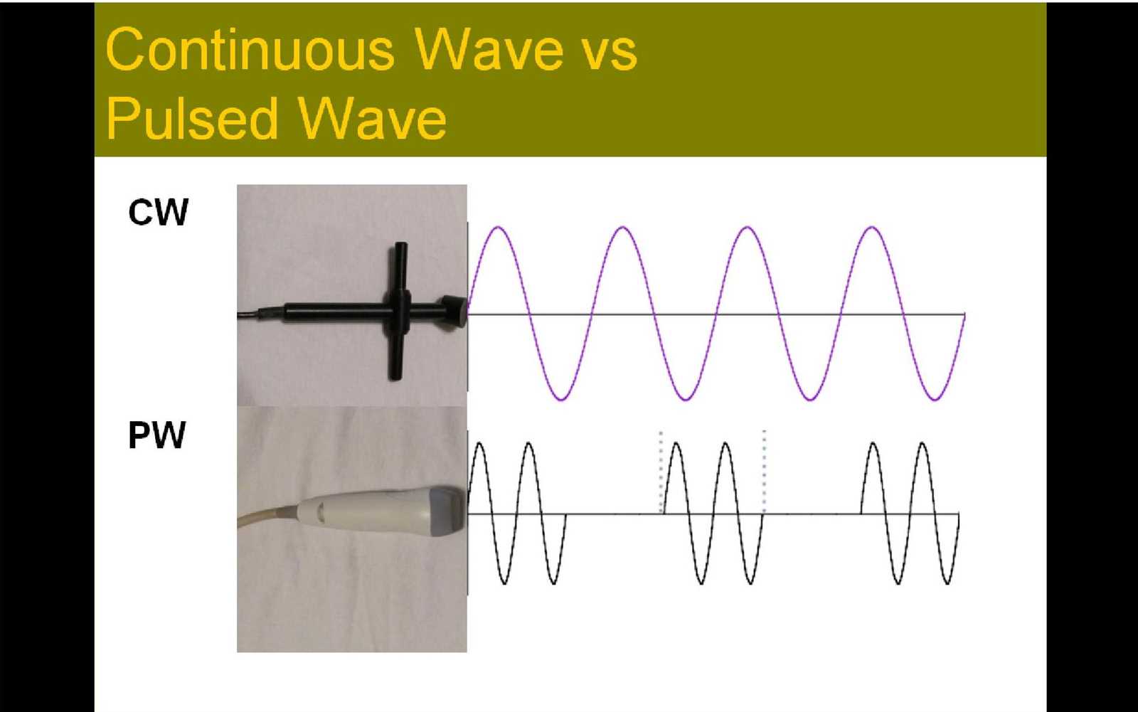
In the realm of diagnostic technology, a deep understanding of the fundamental principles behind sound waves and their interaction with tissues is crucial. The ability to interpret these principles ensures accuracy and efficiency in medical evaluations. Grasping the core concepts allows practitioners to confidently analyze images and make informed decisions during assessments.
Concepts such as wave behavior, transmission, and signal processing form the backbone of any advanced imaging system. By focusing on these, one can better understand the mechanics that enable non-invasive visualizations of the body. This knowledge is not only essential for academic success but also for practical application in clinical settings.
Preparation for assessments in this field requires a solid foundation in theoretical knowledge, as well as the ability to apply this understanding to solve real-world challenges. A strategic approach to mastering these topics will significantly enhance performance in evaluations and daily practice. Focused study of key principles is the pathway to proficiency and success in this ever-evolving field.
Ultrasound Physics Exam Questions
Mastering the key concepts related to sound wave behavior and their application in medical imaging is essential for success in assessments. The ability to understand how different factors influence image quality and diagnostic accuracy is vital. A deep knowledge of wave transmission, reflection, and attenuation forms the core of this subject and enables professionals to accurately interpret images.
Commonly Tested Principles
In most assessments, the focus is on the foundational principles that govern wave propagation and interaction with tissues. These include the understanding of frequency, wavelength, and how they relate to image resolution. The ability to explain the role of sound speed and its impact on diagnostic outcomes is another critical area. Additionally, understanding how different tissue types affect wave behavior and influence the resulting images is often explored.
Approaching Practical Scenarios
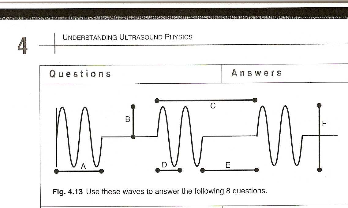
Many scenarios presented in evaluations require applying theoretical knowledge to practical situations. For instance, interpreting how different parameters affect image clarity and resolution. Identifying the correct transducer settings based on specific clinical conditions is a common challenge. Preparing for these situations involves not only knowing the theory but also practicing with realistic case studies that mimic real-world imaging challenges.
Understanding the Basics of Ultrasound Physics
At the heart of diagnostic imaging lies the understanding of how sound waves interact with different materials. By grasping the fundamental principles behind wave propagation, one can better comprehend how images are formed and how these images are used for accurate medical assessment. Sound waves travel through various tissues, and their behavior–such as reflection, refraction, and absorption–determines the quality of the resulting image.
Key concepts include the nature of waveforms, frequency, and wavelength, which directly influence the clarity and resolution of the captured images. Understanding how different tissues, such as muscles, fat, and organs, affect sound wave transmission is critical for accurate diagnosis. These foundational principles not only provide the theoretical framework but also enable the practical application of technology in real-world clinical settings.
Key Concepts in Ultrasound Wave Propagation
Understanding the behavior of sound waves as they travel through various materials is essential for effective imaging. The way these waves interact with different tissues influences the quality of the resulting images. Key principles like wave velocity, frequency, and wavelength are central to this process. These elements determine how sound waves are reflected, refracted, and absorbed by different substances in the body.
Wave velocity is a critical factor in determining how quickly sound waves move through different mediums. This varies depending on the density and elasticity of the material. Similarly, the frequency of the wave plays a significant role in image resolution, with higher frequencies providing greater detail but less penetration. Understanding these concepts allows for optimizing image quality and achieving accurate diagnostics.
Types of Ultrasound Imaging Techniques
Various methods are employed to capture internal images using sound waves, each suited for different diagnostic needs. These techniques rely on distinct approaches to sound wave transmission and reception, offering unique advantages based on the clinical situation. By selecting the most appropriate method, healthcare professionals can obtain the clearest and most accurate images for a wide range of medical conditions.
| Technique | Description |
|---|---|
| 2D Imaging | A basic method producing flat, two-dimensional images of the body’s internal structures, commonly used for routine diagnostic procedures. |
| 3D Imaging | Provides a three-dimensional view, offering more detailed and realistic representations of organs and tissues, often used in obstetrics and cardiology. |
| Doppler Imaging | Measures the movement of fluids (like blood) within the body, allowing the visualization of blood flow and detection of blockages or abnormalities. |
| Elastography | Measures tissue stiffness, helping to identify areas of concern such as tumors or liver fibrosis. |
| Contrast Imaging | Uses contrast agents to enhance the visibility of certain structures or abnormalities, improving image quality in difficult-to-see areas. |
Importance of Frequency in Ultrasound
The frequency of sound waves plays a critical role in determining the quality and effectiveness of imaging techniques. By adjusting the frequency, medical professionals can influence the resolution and depth of penetration of the waves, allowing for better visualization of internal structures. The choice of frequency impacts both the clarity of the images and the ability to detect abnormalities within various tissues.
High Frequency for Detailed Imaging
Higher frequencies provide greater resolution, making it possible to capture finer details of superficial structures. This is especially beneficial for imaging organs close to the surface, such as the thyroid or muscles. However, higher frequencies have lower penetration power, limiting their use for deeper tissues.
Lower Frequency for Deeper Penetration
Lower frequencies, while offering less resolution, have a greater ability to penetrate deeper into the body. This makes them ideal for imaging larger organs or structures that are situated farther from the surface, such as the liver or kidneys. The trade-off is a loss of fine detail, but these frequencies are crucial for comprehensive diagnostic assessments.
Role of Sound Speed in Diagnostics
The speed at which sound waves travel through different tissues is a key factor in diagnostic imaging. This property directly influences how signals are reflected, received, and processed to create clear and accurate images. Variations in sound speed help differentiate between different types of tissues, making it easier to identify abnormalities and assess overall health.
Factors Affecting Sound Speed

Several factors influence the speed at which sound waves travel through the body:
- Tissue Density: Denser tissues, like bone, cause sound waves to travel more slowly, while less dense tissues, such as fat, allow for faster transmission.
- Temperature: Higher temperatures typically result in an increase in sound speed, as the molecules in the medium move more rapidly.
- Medium Type: Different materials, such as water, air, and soft tissue, each have unique properties that affect sound wave velocity.
Impact on Diagnostic Accuracy

Understanding how sound speed varies across different tissues helps improve diagnostic accuracy by providing clearer differentiation between structures. For example, slower sound speeds in organs like the liver allow for more detailed images, while faster speeds in muscle tissue enable quicker data collection. These differences are essential for creating precise, high-quality scans that are crucial for accurate diagnosis and treatment planning.
Attenuation and Its Effect on Images
Attenuation refers to the reduction in intensity of sound waves as they travel through different tissues. This phenomenon occurs due to the absorption, scattering, and reflection of the waves within the body. As sound waves lose energy, their ability to create clear and detailed images diminishes, potentially affecting the diagnostic process.
Factors Contributing to Attenuation
Several factors influence how much attenuation occurs as sound waves pass through tissues:
- Tissue Type: Denser tissues, like bone, cause greater attenuation compared to softer tissues like muscles or fat.
- Frequency: Higher frequencies are more easily attenuated because they have shorter wavelengths and are more prone to scattering and absorption.
- Distance: The greater the distance the sound waves travel, the more they are attenuated, leading to weaker signals and potentially less accurate images.
Impact on Image Quality
As sound waves undergo attenuation, the resulting images can lose clarity and contrast, which may make it more difficult to detect subtle abnormalities. Areas deeper within the body, where waves have traveled a longer distance, are particularly affected. To counteract attenuation, techniques such as adjusting the frequency or using contrast agents can be employed to enhance image quality and diagnostic accuracy.
Resolution and Its Impact on Accuracy
Resolution is a critical factor that determines the level of detail captured in diagnostic imaging. It refers to the ability to distinguish between closely spaced objects or structures. Higher resolution enables clearer and more precise images, which are essential for accurately identifying abnormalities and making correct diagnoses. When resolution is compromised, even significant conditions may go undetected or be misinterpreted.
The accuracy of any diagnostic technique heavily depends on the clarity of the images it produces. If the resolution is too low, small structures may appear blurred or merged, leading to false conclusions. By adjusting the imaging parameters and ensuring optimal resolution, healthcare professionals can improve diagnostic confidence and minimize the risk of errors.
Understanding Doppler Effect in Ultrasound
The Doppler effect plays a significant role in the ability to assess the movement of fluids or tissues within the body. It occurs when there is a shift in the frequency of waves as they reflect off moving objects. This principle allows for the measurement of velocity and flow patterns, which are essential for diagnosing various medical conditions, such as blood flow issues and organ function.
How Doppler Effect Works
The Doppler effect is based on the change in frequency or wavelength of the waves due to motion. This can be understood through the following principles:
- Moving Towards the Source: When the object moves toward the sensor, the frequency of the waves increases, resulting in a higher pitch or frequency shift.
- Moving Away from the Source: When the object moves away, the frequency decreases, producing a lower pitch or frequency shift.
- Measurement of Velocity: The amount of frequency shift is directly proportional to the speed of the moving object, allowing for accurate velocity measurements.
Applications in Diagnostics
By analyzing these frequency shifts, medical professionals can gain crucial insights into the movement and condition of blood vessels, heart valves, and other organs. The Doppler effect is widely used in diagnostics to detect abnormalities such as blockages, blood clots, and irregular blood flow, enhancing overall diagnostic accuracy and patient care.
Analyzing Pulse Echo and its Uses
The pulse echo technique is a fundamental principle in imaging systems that rely on sound waves to detect structures within the body. It involves sending short bursts of sound energy into tissues and then measuring the time it takes for the echoes to return. The reflection of these pulses helps to create an image of the internal structures, allowing for a detailed assessment of various organs and tissues.
By analyzing the time it takes for sound waves to travel to a target and return, the system can calculate the distance to the object, helping to map the internal environment accurately. This method is essential for determining the location, size, and characteristics of objects inside the body, which is invaluable for diagnosis and treatment planning.
Transducer Types and Their Functionality
Transducers are key components in imaging systems that convert energy from one form to another, allowing the detection of internal structures through sound waves. These devices play a vital role in capturing and transmitting signals, enabling the creation of accurate images. Different types of transducers are designed to meet specific diagnostic needs, each with its own unique capabilities and applications.
Common Transducer Types
There are several common transducer types, each suited for particular imaging requirements:
- Linear Array: This transducer consists of a series of small elements arranged in a straight line, ideal for imaging superficial structures and producing high-resolution images.
- Curved Array: With a curved arrangement of elements, this transducer is designed for deeper imaging, offering a wider field of view while maintaining good resolution at various depths.
- Phased Array: These transducers are compact and versatile, commonly used in cardiac imaging, as they can focus on a specific area from different angles with excellent depth penetration.
Functionality and Applications
Each type of transducer has distinct advantages depending on the clinical situation. For instance, a linear array is typically used for imaging soft tissues near the surface, such as muscles or tendons, while a phased array is preferred for visualizing deeper structures, such as the heart or large blood vessels. Understanding the specific functionality of each transducer helps healthcare professionals choose the right tool for accurate diagnostics and effective treatment planning.
Physics Behind Reflection and Refraction
The interaction of sound waves with different tissues and materials plays a crucial role in creating diagnostic images. Two fundamental processes that occur when sound waves travel through various mediums are reflection and refraction. These phenomena are essential for visualizing internal structures and accurately assessing the body’s condition.
When sound waves encounter a boundary between two different materials, part of the energy is reflected back while the rest continues to travel through the medium. The amount of reflection depends on the differences in acoustic properties between the materials. Refraction occurs when sound waves change direction as they pass through different mediums, altering their speed. This bending of waves can distort the image if not properly accounted for.
Key Concepts in Reflection
- Angle of Incidence: The angle at which the sound waves strike a surface determines the angle of reflection. If the angle is too steep, more energy is reflected back, reducing the amount of sound that penetrates deeper tissues.
- Impedance Mismatch: The greater the difference in acoustic impedance between two materials, the stronger the reflection. This principle is used to distinguish between different tissues, such as between muscles and fat.
Key Concepts in Refraction
- Change in Speed: When sound waves pass from one medium to another with a different speed of sound, they bend. This effect is especially significant in tissues with varying densities, such as between soft tissue and bone.
- Critical Angle: If sound waves strike a medium boundary at a specific angle, total internal reflection can occur, meaning no energy will pass through. This phenomenon can limit the depth of penetration in certain diagnostic procedures.
By understanding how sound waves behave through reflection and refraction, medical professionals can optimize imaging techniques, ensuring clearer and more accurate results. These principles are fundamental to the effectiveness of diagnostic tools in detecting and evaluating internal conditions.
Safety Considerations in Ultrasound Exams
Ensuring safety during diagnostic procedures is of utmost importance, especially when using technologies that involve the use of sound waves for imaging. While these methods are generally non-invasive and considered safe, it is essential to follow proper protocols to minimize any risks to patients and operators. Understanding the key safety considerations helps to maintain the integrity of the process and avoid potential hazards.
Key factors that influence safety include the frequency and intensity of sound waves, as well as the duration of exposure. In medical applications, these variables are carefully regulated to ensure that the procedure does not cause harm while providing accurate diagnostic results. Below are some key safety practices to consider:
Frequency and Intensity Management
- Appropriate Frequency Range: Using the right frequency ensures optimal image quality while minimizing any risks associated with sound wave penetration. Higher frequencies provide better resolution but have limited tissue penetration, while lower frequencies penetrate deeper but may offer less detail.
- Controlled Power Output: The power of the sound waves should be adjusted to the minimum level required for accurate imaging. Excessive energy exposure can lead to thermal effects or tissue damage.
Duration and Repetition of Exposure
- Minimizing Exposure Time: Limiting the duration of each session helps to reduce the risk of heat generation and potential harm to the tissues. It is important to balance the need for quality imaging with the duration of exposure.
- Avoiding Unnecessary Repeated Procedures: Repeated exposures, even at low levels, can accumulate and potentially cause long-term effects. It’s crucial to only conduct tests when absolutely necessary for diagnosis or treatment.
Adhering to these guidelines ensures that diagnostic procedures remain safe, effective, and reliable, providing accurate results while protecting the well-being of patients. When performed by trained professionals and under regulated conditions, these imaging techniques offer significant benefits with minimal risk.
Common Mistakes in Ultrasound Physics Exams
When preparing for assessments involving diagnostic imaging technology, candidates often make several recurring mistakes that can hinder their performance. These errors, while common, can be easily avoided with proper understanding and preparation. Recognizing these pitfalls is the first step in improving accuracy and achieving better results. This section highlights some of the most frequent errors students make and how to overcome them.
Misunderstanding Key Concepts
- Confusing Terminology: One of the most frequent mistakes is mixing up terms that appear similar but have different meanings. It’s crucial to understand the specific definitions and applications of terms to avoid confusion during assessments.
- Lack of Conceptual Clarity: Without a clear understanding of the fundamental principles behind the techniques, students may misinterpret questions or fail to apply concepts correctly. Thoroughly reviewing basic principles and practicing application scenarios is vital.
Poor Time Management
- Rushing Through Questions: Some candidates attempt to answer questions too quickly, especially in high-pressure situations. This leads to careless errors. Proper time management allows for thoughtful consideration of each question, reducing the risk of mistakes.
- Skipping Over Complex Questions: In an attempt to save time, students sometimes skip difficult questions only to return to them later when they’ve run out of time. It’s better to approach each question methodically and allocate time based on the difficulty level of the question.
By recognizing these common errors and taking proactive steps to address them, candidates can improve their exam performance. Focusing on the details, practicing regularly, and staying organized can significantly enhance one’s understanding and test-taking ability.
Preparing for Ultrasound Physics Questions
Effective preparation for assessments related to diagnostic imaging technologies involves not just studying theory but also honing practical skills and critical thinking. Understanding the fundamental principles is essential, but applying them in various scenarios can make the difference between a good and great performance. This section outlines strategies and approaches that can help in efficiently preparing for these assessments.
Mastering Key Concepts
To excel in these assessments, it is important to have a strong grasp of core principles. Focus on understanding the fundamental theories behind the techniques, including wave propagation, energy transfer, and signal interpretation. It’s not enough to memorize formulas; understanding their practical application is essential. The following strategies can help:
- Break down complex concepts into smaller, manageable chunks.
- Use visual aids and diagrams to reinforce understanding.
- Practice applying concepts to real-world scenarios.
Practical Training and Mock Tests
Hands-on practice is crucial to mastering the material. Engaging in mock tests can simulate the exam environment, helping to build confidence and reduce anxiety. Moreover, mock tests often reveal areas that need improvement. It’s advisable to:
- Set a timer and practice answering questions under timed conditions.
- Review past tests and identify recurring question types and patterns.
- Seek feedback from peers or instructors to refine your approach.
By following these preparation strategies, students can build a comprehensive understanding and boost their chances of success in their assessments. A well-rounded approach that combines theory, practice, and mock tests will ensure readiness and confidence when tackling these challenging topics.
Practice Questions for Ultrasound Physics Exam
To effectively prepare for assessments on diagnostic imaging principles, it’s important to engage with practice material that mirrors the types of challenges encountered during tests. Practicing with sample scenarios allows for reinforcing knowledge, improving problem-solving skills, and boosting confidence. This section offers a selection of practice exercises to help assess your understanding of the key concepts involved in these technical assessments.
Sample Problems for Skill Building
Here are a few practice problems that cover essential topics in the field. They are designed to test both theoretical knowledge and practical application.
| Problem | Answer | Explanation |
|---|---|---|
| What happens to the intensity of a wave as it travels through different mediums? | It decreases due to absorption and scattering. | As waves move through various materials, energy is lost because of absorption and scattering, which reduces their intensity. |
| How does the speed of sound change with temperature? | The speed increases as temperature rises. | Temperature affects the speed of sound; as the temperature increases, the molecules in the medium move faster, allowing sound waves to travel more quickly. |
| What is the relationship between wavelength and frequency? | Wavelength is inversely proportional to frequency. | As the frequency of a wave increases, its wavelength decreases, and vice versa, following the equation: λ = v / f. |
Advanced Problem Set for Practice
The following set of more advanced problems will challenge your comprehension of the material in more complex scenarios:
| Problem | Answer | Explanation |
|---|---|---|
| If the intensity of a wave is halved, what happens to the power? | The power is also halved. | Intensity and power are directly related, so halving one results in halving the other. |
| How do you calculate the impedance of a medium? | Impedance = density × speed of sound. | Impedance is calculated by multiplying the density of the medium by the speed at which sound waves travel through it. |
| What effect does increasing frequency have on resolution? | Resolution improves, but penetration decreases. | Higher frequencies provide better resolution due to smaller wavelengths, but they also result in poorer penetration into tissues. |
Engaging with these problems will help reinforce key concepts and enhance problem-solving abilities, giving you the confidence to tackle similar questions in an assessment environment.
Advanced Topics in Ultrasound Physics
In the realm of diagnostic imaging, there are several advanced principles and techniques that go beyond the foundational concepts. These specialized topics require a deeper understanding of wave behavior, signal processing, and the intricacies of how sound waves interact with various tissues. This section delves into the more complex aspects of these technologies, offering insights into cutting-edge methods used in clinical and research settings.
Wave Interactions and Signal Processing
One of the most critical areas of advanced study is the way sound waves interact with different types of tissue and how these interactions are processed to create accurate images. A thorough understanding of reflection, refraction, and scattering is essential for improving diagnostic quality and precision.
- Reflection and Refraction: These phenomena occur when sound waves encounter boundaries between different tissue types. Reflection is vital for producing high-quality images, while refraction can affect image clarity and lead to artifacts.
- Scattering: As sound waves travel through the body, they encounter various structures that scatter the waves, influencing the final image. This can be particularly important in evaluating tissue types like muscle, bone, or fat.
- Signal Processing: After waves are reflected or scattered, they need to be processed to generate usable data. Advanced algorithms and filtering techniques are often employed to enhance image quality and resolve finer details.
Advanced Imaging Techniques and Applications
There are also numerous sophisticated imaging methods that push the limits of conventional diagnostic imaging. These techniques are designed to improve accuracy, image resolution, and even enable real-time imaging in dynamic environments.
- Doppler Imaging: This technique is used to measure the velocity of blood flow and tissue movement by analyzing the frequency shift of sound waves. It is particularly valuable in cardiology and vascular imaging.
- Harmonic Imaging: By utilizing higher-frequency sound waves, harmonic imaging can provide clearer images with better contrast, particularly in deeper tissues.
- Elastography: This method evaluates tissue stiffness by measuring the propagation of shear waves, providing valuable information for assessing liver fibrosis, tumors, and other conditions.
Mastering these advanced topics is essential for professionals aiming to push the boundaries of diagnostic imaging and utilize the latest innovations for improved patient outcomes.
Exam Strategies for Ultrasound Physics
Successfully tackling assessments in diagnostic imaging requires more than just knowledge of core concepts; it demands effective strategies to navigate complex material and maximize performance. Preparing for these assessments involves a blend of understanding theory, applying practical knowledge, and mastering time management. This section highlights key approaches that can help boost success when facing challenging assessments related to sound wave behavior, imaging techniques, and associated technologies.
Time Management and Question Approach
One of the most crucial elements of preparation is managing your time effectively during the test. Here’s how you can approach the questions with confidence:
- Prioritize Easy Questions: Start with the questions you are most confident in. This allows you to secure quick points and build momentum.
- Allocate Time for Each Section: Break down your available time according to the number of sections or questions. Make sure to leave time for reviewing and double-checking your answers.
- Don’t Get Stuck: If a question is proving difficult, move on and come back to it later. Spending too much time on one question can cause you to run out of time for others.
Understanding Common Topics and Concepts
Familiarity with the most commonly tested topics can significantly reduce the time spent on recalling information. Focus on the following areas:
| Topic | Key Focus |
|---|---|
| Wave Propagation | Understand how sound waves travel through different mediums, including reflection, refraction, and attenuation. |
| Signal Processing | Review how signals are processed to form images, including filtering, amplification, and noise reduction. |
| Imaging Techniques | Learn the differences between various techniques such as Doppler, harmonic, and elastography, and their specific applications. |
| Safety Protocols | Be sure to understand the safety guidelines and the principles behind non-invasive testing. |
By focusing on these areas, you can ensure that you are well-prepared for the assessment and able to approach each question methodically. Remember, practice and familiarity with the material are key to improving both your understanding and test performance.