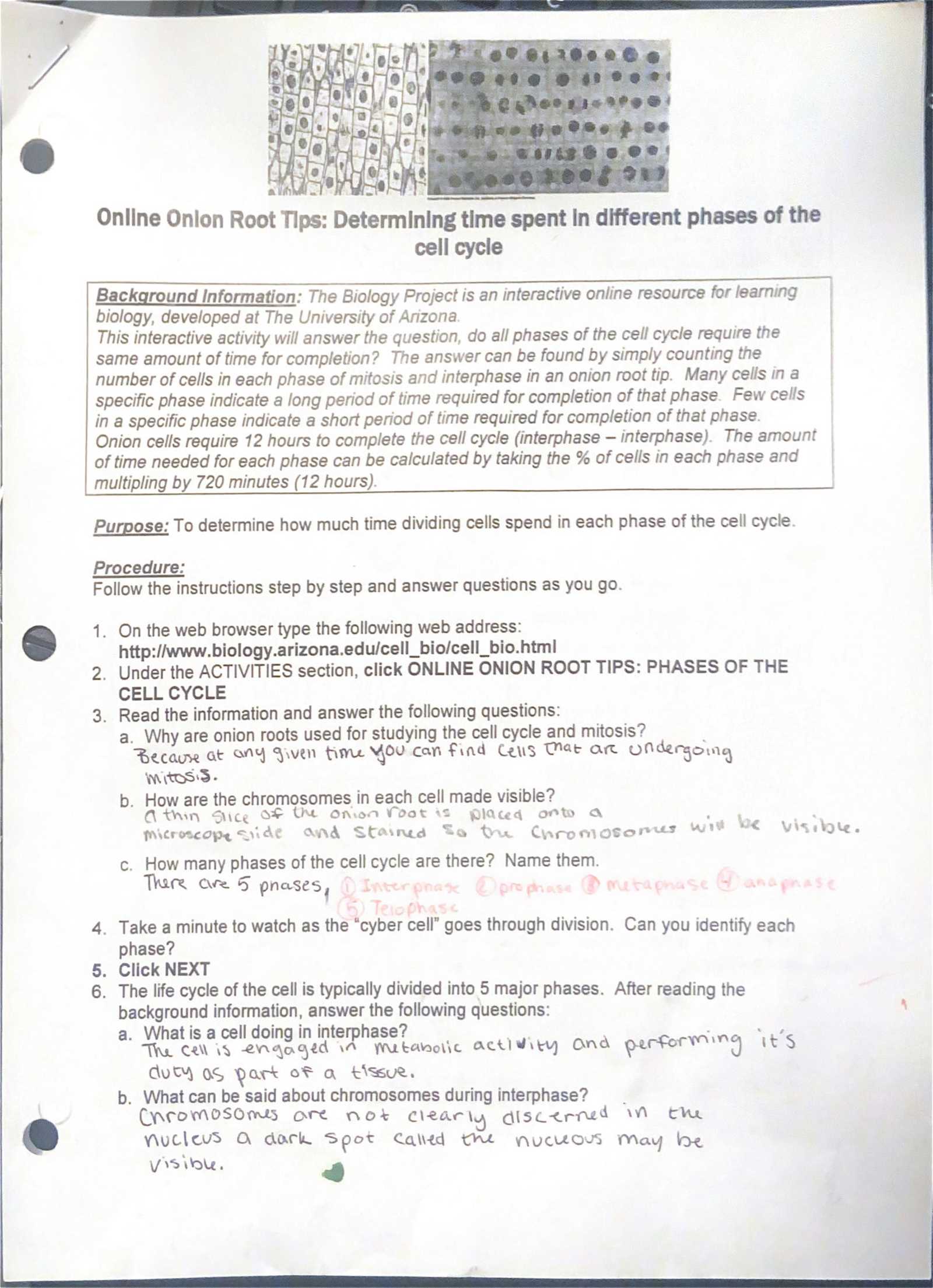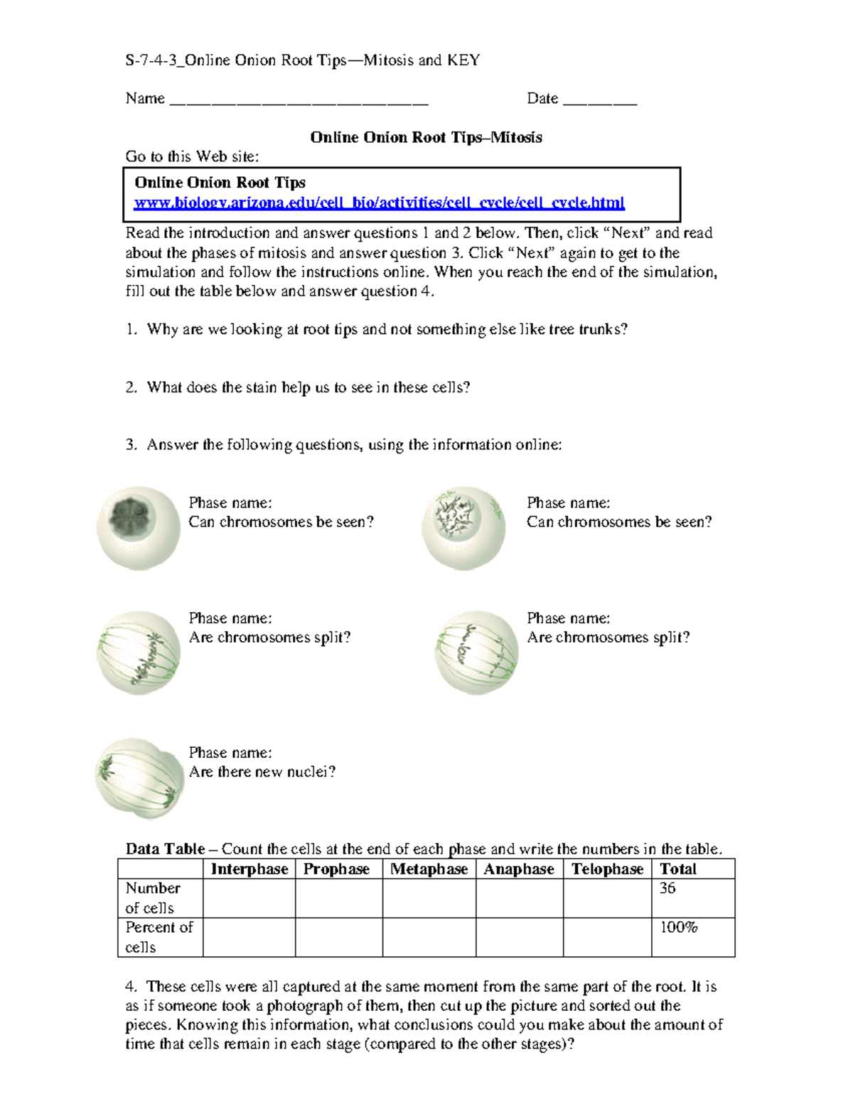
Studying the process of cell division is a fundamental aspect of biology education. By examining the growth stages of plants, students can observe and analyze how cells divide and multiply. This process, known as mitosis, is essential for the development of new tissues and organisms. In particular, certain plant cells provide a clear view of this process, making them an ideal choice for laboratory analysis.
Through hands-on experimentation, learners can explore the stages of mitosis and understand how cells transition from one phase to the next. The small and fast-growing plant cells used in these studies allow for easy observation under a microscope. By studying how cells divide, students gain insights into larger biological processes, from tissue regeneration to organismal growth.
Proper observation and analysis of cell division in plants provide students with an opportunity to enhance their critical thinking skills. Understanding the details of this process contributes to a deeper grasp of genetics, cellular biology, and the mechanics of life itself. With accurate interpretations of these studies, students can develop a clearer understanding of biological systems and their complexities.
Understanding Cell Division in Plant Experiments
In biological studies, examining cell division through plant specimens offers a valuable opportunity to explore the mechanisms of growth and replication. By observing certain plant cells under a microscope, students can gain insights into how individual cells undergo division and contribute to the development of tissues. This hands-on approach allows for a deeper understanding of the phases of mitosis and how they unfold in real-time.
Observing the Stages of Mitosis
During the process of cell division, plants enter distinct stages that can be observed through microscopic analysis. These stages, such as prophase, metaphase, anaphase, and telophase, represent the steps through which the cell’s genetic material is replicated and distributed to new cells. By carefully analyzing plant cells during these stages, learners can gain clarity on how cellular structures behave during mitosis.
Interpreting Results and Learning Outcomes
Accurate interpretation of these observations leads to a better understanding of the genetic and cellular processes at play. Calculating the frequency of each phase and identifying abnormalities allows students to connect theoretical knowledge with practical application. This method of exploration enhances critical thinking and provides a more thorough understanding of biological functions in living organisms.
Understanding the Plant Cell Division Experiment
In biological studies, observing the division of cells through plant material offers an effective way to learn about cellular processes. By focusing on specific plant structures, researchers can study how cells replicate and multiply to form new tissue. This experiment serves as an introduction to key concepts like mitosis and the cell cycle, making it a valuable educational tool for understanding basic biological principles.
Preparing Plant Material for Observation
To observe cell division, certain plant sections are carefully selected for their ability to display rapid growth and cell activity. These sections are prepared and stained to enhance visibility under a microscope, allowing students to clearly see the stages of mitosis. The ease with which these specimens can be observed makes them an ideal choice for learning about cellular processes in a controlled environment.
Key Concepts in Cell Division
Understanding the stages of cell division–such as the replication of DNA, alignment of chromosomes, and the separation of genetic material–is essential in grasping how organisms grow and develop. The experiment helps students visualize these stages in action, offering a clearer understanding of how cell division supports tissue formation and regeneration in living organisms.
Steps to Complete the Plant Cell Division Experiment
To successfully observe cell division in plant specimens, a series of steps must be followed to ensure clear results. These steps include preparing the material, staining the cells, and carefully observing the process under a microscope. By following the proper procedure, students can gain a deeper understanding of how cells replicate and divide during growth.
| Step | Description |
|---|---|
| 1. Select Plant Sections | Choose fast-growing plant parts, typically near the tips, where cell division is most active. |
| 2. Prepare the Specimen | Cut the plant material into small pieces for easier observation under the microscope. |
| 3. Stain the Cells | Apply a staining solution to make the chromosomes more visible and enhance contrast. |
| 4. Mount the Specimen | Place the prepared sample onto a microscope slide with a coverslip. |
| 5. Observe Under the Microscope | Examine the specimen at different magnifications to observe various stages of cell division. |
| 6. Record the Findings | Document the observed stages of cell division and calculate relevant data, such as mitotic index. |
Key Concepts in Mitosis for Students
Mitosis is a critical process in cell biology, responsible for growth, tissue repair, and asexual reproduction. Understanding the stages of this process is essential for students studying biology, as it explains how organisms develop and maintain their cellular structures. By examining how a single cell divides to form two genetically identical daughter cells, students can grasp the fundamental mechanisms of cellular function and reproduction.
- Interphase: The cell prepares for division by replicating its DNA and increasing its size. This phase consists of G1, S, and G2 stages.
- Prophase: The chromatin condenses into visible chromosomes, and the nuclear membrane begins to break down.
- Metaphase: Chromosomes align at the center of the cell, attached to spindle fibers that guide their movement.
- Anaphase: Sister chromatids are pulled apart toward opposite ends of the cell, ensuring each daughter cell receives a full set of chromosomes.
- Telophase: The nuclear membrane reforms around the separated chromosomes, which begin to de-condense.
- Cytokinesis: The cytoplasm divides, completing the formation of two separate daughter cells.
These stages are vital to understanding how genetic material is accurately distributed and how cells contribute to the development and maintenance of multicellular organisms. By mastering these core concepts, students can gain a deeper appreciation for the complexity of life at the cellular level.
How to Prepare Plant Sections for Study
Preparing plant specimens for microscopic examination involves a series of steps designed to highlight the stages of cell division. The process ensures that the cells are visible and their structures can be clearly observed. By following these steps, you can effectively prepare the material for study, allowing for accurate observation of mitosis and other cellular activities.
Start by selecting the appropriate plant sections, typically from fast-growing areas where cell division is most active. These parts of the plant contain cells that are in various stages of division, making them ideal for observation. Once the material is selected, it must be carefully cut into small pieces, ensuring that the cells remain intact and undamaged.
Next, the specimens are treated with a staining solution to enhance the contrast and make the chromosomes more visible under the microscope. The staining helps differentiate various cellular components, such as the nucleus, chromosomes, and spindle fibers. After staining, the prepared sections are mounted on a microscope slide and covered with a coverslip, making sure there are no air bubbles that could interfere with the view.
Finally, once the specimen is prepared, it is ready for observation under the microscope. Adjusting the focus and magnification will allow you to observe the various stages of mitosis, from prophase to cytokinesis, providing valuable insights into the process of cell division.
Importance of Plant Sections in Science
Plant specimens, particularly those with rapidly dividing cells, play a crucial role in advancing our understanding of cellular processes. These materials offer clear and easily observable stages of cell division, making them valuable for studies in biology. Their consistent growth patterns and transparent nature allow scientists and students alike to examine cellular activities in detail, providing insight into the fundamental mechanisms of life.
One of the primary reasons these plant sections are widely used in scientific research is their ability to demonstrate the stages of mitosis–the process through which cells replicate. The cells in these plants divide rapidly, making them ideal for observing the progression from one phase of mitosis to the next. By studying these processes in plant cells, scientists can apply their findings to broader concepts of cellular biology, genetics, and even cancer research, where uncontrolled cell division plays a role.
In addition, these plant sections are often used in educational settings, helping students visualize complex biological concepts in a hands-on environment. The clear visibility of chromosomes, cell structures, and other key components during division enhances learning and fosters a deeper understanding of how organisms grow and develop. The simplicity and accessibility of using plants for such studies make them indispensable tools in both academic and scientific settings.
Common Mistakes in Plant Cell Division Experiment
When conducting an experiment to observe cell division in plant specimens, there are several common errors that can affect the results. These mistakes often arise from improper preparation, handling, or observation of the plant material. Understanding and avoiding these errors is crucial to obtaining accurate and reliable data. Below are some of the most frequently encountered mistakes in this type of experiment.
Incorrect Preparation of Specimens
- Improper Cutting: Cutting the plant material too thick or unevenly can make it difficult to observe individual cells under the microscope.
- Inadequate Staining: Using too little or too much staining solution can obscure important details in the cell structure, making it difficult to differentiate stages of division.
- Rough Handling: Handling the specimens too roughly can damage the delicate cells, leading to inaccurate or incomplete observations.
Microscope Handling Errors
- Incorrect Focusing: Failing to properly adjust the focus can result in blurry images, making it challenging to clearly identify the stages of cell division.
- Improper Magnification: Using too high or too low a magnification can prevent you from seeing the necessary details, either zooming in too much or missing key structures entirely.
By being mindful of these common mistakes and taking extra care during the experiment, you can improve the accuracy of your observations and better understand the stages of cell division in plants.
Observing Cell Division in Plant Sections
Studying the process of cell division in plant specimens provides valuable insights into how organisms grow and develop. By observing the different stages of mitosis, researchers can learn about cellular structures and the mechanisms that ensure accurate genetic replication. The plant sections chosen for this purpose are ideal due to their active growth and the ease with which their cells can be examined under a microscope.
The procedure for observing cell division in plant sections involves several key steps to ensure that the stages of mitosis are clearly visible. The process requires careful preparation of the specimen, proper staining to enhance contrast, and the correct use of a microscope to examine the cells in detail. The following are some important stages and observations that can be made during the experiment:
- Prophase: Chromosomes become visible, and the nuclear membrane begins to break down.
- Metaphase: Chromosomes align at the center of the cell, preparing for division.
- Anaphase: Chromatids are pulled apart toward opposite poles of the cell.
- Telophase: New nuclear membranes form around the separated chromatids, marking the final stages of division.
- Cytokinesis: The cytoplasm divides, completing the process of cell division and resulting in two daughter cells.
By following the correct procedures and carefully observing these stages, students and researchers can gain a deeper understanding of the vital processes that support life at the cellular level.
Mitotic Stages in Plant Cells
Understanding the stages of cell division is essential for studying growth and development in organisms. In plant cells, the process of mitosis is vital for generating new cells that contribute to tissue growth. The division of cells occurs in a sequence of distinct phases, each characterized by specific changes in the cell’s structure. Observing these stages helps in understanding the precision and complexity involved in cellular reproduction.
The following table outlines the key stages of mitosis observed in plant cells, particularly those from rapidly dividing growth zones. Each phase of the process ensures that the genetic material is properly replicated and distributed between two daughter cells:
| Stage | Description |
|---|---|
| Prophase | The chromosomes condense and become visible under the microscope. The nuclear membrane starts to break down. |
| Metaphase | Chromosomes align along the middle of the cell, attached to spindle fibers that guide their movement. |
| Anaphase | The sister chromatids are pulled apart toward opposite poles of the cell, ensuring each new cell gets an identical set of chromosomes. |
| Telophase | The chromosomes begin to de-condense, and new nuclear membranes form around each set of chromosomes. |
| Cytokinesis | The cytoplasm divides, resulting in the formation of two distinct daughter cells. |
By examining these stages in plant cells, researchers and students can better understand the process of cell division and its role in growth and development.
Calculating Mitotic Index in Plant Cells
The mitotic index is a valuable tool used to measure the rate of cell division within a given tissue sample. This index is calculated by determining the proportion of cells undergoing mitosis at any given time, which reflects the activity of growth and regeneration processes in an organism. By calculating the mitotic index in plant specimens, researchers can assess the health and growth rate of plant tissues, especially in rapidly dividing regions.
Steps to Calculate Mitotic Index
To calculate the mitotic index, follow these steps:
- Prepare the Sample: Select a region of the plant where cell division is actively occurring. Prepare the sample by properly staining the cells to enhance visibility under a microscope.
- Count Total Cells: Under the microscope, count the total number of cells in the prepared sample. This gives you the total cell population in the field of view.
- Identify Mitotic Cells: Identify and count the number of cells in the different stages of mitosis (prophase, metaphase, anaphase, telophase).
- Calculate the Index: Divide the number of cells in mitosis by the total number of cells observed and multiply by 100 to get the mitotic index percentage.
Interpreting the Results

A higher mitotic index indicates a higher rate of cell division, often seen in tissues undergoing rapid growth. Conversely, a lower index suggests slower cell division, which may occur in mature tissues or during periods of growth arrest. The mitotic index is an important measure in studies related to plant development, genetic research, and cancer biology, where cell division is a central theme.
Why Use Plant Roots in Experiments
Plant roots are often used in scientific experiments due to their unique characteristics and accessibility. These structures are highly active in terms of cellular division and growth, making them ideal for studying processes such as mitosis and cellular development. The root tips of many plants provide a concentrated area where cell division is occurring at a rapid rate, allowing for detailed observation under a microscope.
Advantages of Using Plant Roots
There are several reasons why plant roots are chosen for educational and research purposes:
- Ease of Access: Plant roots are readily available and can be easily collected from commonly grown plants such as garlic, beans, and other fast-growing species.
- Visible Cell Division: The tips of plant roots contain regions where cells divide rapidly, providing a clear view of various stages of mitosis, which is essential for studying cellular processes.
- Non-Destructive Sampling: Harvesting root tips does not harm the overall plant, making it a non-invasive method for research and teaching.
Educational Benefits
Using plant roots for experiments also offers significant educational value. Students can visually understand the different stages of cell division and better grasp concepts such as growth, differentiation, and genetic replication. These hands-on experiments are an effective way to reinforce theoretical knowledge and stimulate interest in biological sciences.
Microscope Techniques for Root Tip Analysis
Effective analysis of plant growth and cell division requires precise microscopy techniques. The study of cellular structures, particularly in actively dividing areas of a plant, can reveal vital information about processes such as mitosis and differentiation. A variety of microscopy methods are used to enhance the visibility of cellular components and ensure accurate observations.
Essential Microscope Techniques

To successfully observe and analyze plant cells, certain techniques are crucial for ensuring high-quality results:
- Proper Staining: Staining the specimen is an essential step to highlight specific cellular structures, making the division process more visible under the microscope.
- Use of High Magnification: High-powered objectives are necessary to view individual cells and their stages in detail, especially for observing mitosis in small regions like growth tips.
- Preparation of Thin Sections: Preparing thin slices or sections of the plant tissue allows light to pass through and improves the clarity of cellular details.
- Use of Contrast Techniques: Adjusting contrast and light settings helps differentiate between various cellular components, making it easier to identify stages of division.
Tips for Accurate Observation
To improve the accuracy and clarity of observations, consider the following tips:
- Focus on the Most Active Areas: Target the tips where cell division is most active for clearer results.
- Ensure Stable Illumination: Proper lighting is key to reducing shadows and enhancing visibility of fine structures.
- Monitor for Motion: Watch for changes in the cells over time to track the progression of division stages.
Analyzing Results from Root Cell Division Experiment
After conducting an experiment on plant cell division, it is crucial to systematically analyze the results to understand the efficiency and patterns of cellular processes. Proper analysis helps identify key stages in the cell cycle, recognize patterns of mitosis, and assess how external factors may influence cellular growth. The following guidelines can help you interpret the findings from the experiment and draw meaningful conclusions.
Key Steps in Data Analysis
To ensure accurate analysis of the experiment, follow these essential steps:
- Count Dividing Cells: Begin by counting the number of cells in different stages of division, such as prophase, metaphase, anaphase, and telophase.
- Calculate Mitotic Index: This ratio of dividing cells to total cells gives insight into the activity level of mitosis within the sample.
- Compare with Controls: If control samples are available, compare the results to understand the impact of various conditions or treatments on cell division.
- Record Observations: Note any variations in cell size, shape, and structure across different stages to detect any anomalies or patterns.
Interpreting the Data
Once the data is collected, interpreting it correctly is vital for understanding cell behavior and growth dynamics:
- Identify Trends: Look for trends in the number of cells in each phase and how they correlate with external factors such as temperature or chemical treatment.
- Understand Mitosis Rates: A higher mitotic index indicates a greater rate of cell division, which may signal faster plant growth or stress responses.
- Report Findings: Summarize the findings in a clear and structured way, highlighting any significant differences or unexpected results.
Lab Safety Tips for Microscopic Studies
When conducting microscopic studies, ensuring a safe environment is essential to protect both the researcher and the equipment. Understanding the proper protocols can prevent accidents and ensure the reliability of the results. By following these safety measures, students and professionals alike can create an optimal environment for study and research.
General Safety Guidelines
Before starting any microscopic investigation, it’s important to follow these general safety protocols:
- Wear Protective Gear: Always wear safety goggles and gloves to prevent exposure to chemicals or potential contamination from specimens.
- Handle Glassware Carefully: Microscopes and slides are often made of glass. Always handle them with care to avoid breakage and potential injury.
- Properly Dispose of Samples: Ensure that any biological or chemical samples are disposed of in designated containers to avoid contamination or harm.
- Keep Work Area Clean: A tidy workspace prevents accidents and ensures that all necessary tools are within easy reach.
Microscope-Specific Safety Tips
In addition to general safety practices, special attention should be given to the use of microscopes:
- Handle with Care: Microscopes are sensitive instruments. Always move and adjust them gently to avoid damage.
- Adjust Light Source Safely: When adjusting the light source, make sure not to stare directly into the light to prevent eye strain or injury.
- Clean Lenses Properly: Use lens paper and a gentle touch to clean the microscope lenses. Avoid harsh chemicals that could damage the optics.
- Never Force the Focus: If the view is blurry, adjust the focus slowly. Forcing the knobs may cause strain on the equipment.
Understanding Chromosomes in Root Cells
Chromosomes play a crucial role in the genetic material of cells, acting as carriers of the hereditary information needed for cell division and function. They are essential for the transmission of traits from one generation to the next and are involved in many cellular processes, including growth, development, and reproduction. In plant cells, observing chromosomes provides valuable insight into the mechanisms of mitosis and genetic variation.
Chromosomes and Their Structure
Chromosomes consist of tightly coiled DNA molecules that are organized into structures visible under a microscope during cell division. Each chromosome is made up of two sister chromatids connected at a region called the centromere. These chromatids contain the genetic code that is crucial for cell functions. The structural organization of chromosomes allows for the accurate distribution of genetic material during mitosis.
Chromosome Behavior During Cell Division
During the process of cell division, chromosomes undergo several stages of alignment and separation to ensure the proper distribution of genetic material. In plant cells, this process can be observed under a microscope, offering insights into the various phases of mitosis, such as prophase, metaphase, anaphase, and telophase. By studying these stages, scientists can understand how cells maintain genetic stability and how errors in this process might lead to mutations or diseases.
Link Between Onion Root Tips and Genetics
Understanding the genetic foundations of cellular processes is a fundamental aspect of modern biology. One of the ways scientists can explore genetics in action is by examining how cells divide and replicate. This process, known as mitosis, is crucial for the growth and development of living organisms. Certain plant cells, particularly those in the tips of growing structures, offer a unique window into this process, revealing key aspects of genetic inheritance and cellular function.
The study of cell division in plants provides an ideal opportunity to observe how genetic material is passed on during cell reproduction. By examining the division of cells in these regions, scientists can better understand how chromosomes behave, align, and segregate during mitosis, which is essential for genetic stability and the transmission of hereditary traits.
Genetic Material in Dividing Cells
The division of cells involves the careful replication and distribution of genetic material, ensuring that each daughter cell inherits the correct set of chromosomes. Through observing mitotic processes in plant cells, it is possible to track how chromosomes are duplicated and segregated, providing insight into genetic processes like genetic variation, mutation, and inheritance. The study of these cells helps scientists understand the basic principles of genetics, such as how traits are inherited across generations.
Table: Stages of Mitosis and Chromosome Behavior
| Stage | Chromosome Behavior |
|---|---|
| Prophase | Chromosomes condense and become visible. The nuclear membrane starts to break down. |
| Metaphase | Chromosomes align at the cell’s equator, attached to spindle fibers. |
| Anaphase | Sister chromatids are pulled apart to opposite sides of the cell. |
| Telophase | New nuclear membranes form around the separated chromosomes. |
This process not only shows how cells replicate but also helps illustrate the relationship between genetics and cellular behavior. By examining the division of cells in these growing structures, we gain valuable insights into how genetic traits are passed on, ensuring the stability and diversity of plant life across generations.
How to Interpret Lab Findings Accurately

Accurate interpretation of experimental data is essential for drawing meaningful conclusions and advancing scientific understanding. When working with biological specimens or conducting cell-based experiments, recognizing key patterns, differentiating variables, and understanding the underlying processes are critical to ensuring reliable results. The ability to analyze and interpret findings correctly helps avoid misinterpretation and supports the validity of scientific claims.
Key Steps for Effective Data Interpretation
When examining experimental outcomes, the following steps can help ensure that the findings are interpreted with precision:
- Ensure Proper Methodology: Use the correct techniques and follow standard procedures. Deviations can lead to inaccurate observations and conclusions.
- Review All Variables: Examine all factors involved in the experiment, including external conditions and possible contaminants, to ensure that results are not skewed by unforeseen influences.
- Validate Findings with Control Samples: Cross-reference results with control samples to confirm that the observed changes are due to the experimental conditions and not other factors.
- Quantify Data: Whenever possible, use numerical data to support qualitative observations. Accurate measurements help reduce subjective biases.
- Consider Statistical Analysis: Apply appropriate statistical tests to determine the significance of your results. Statistical validation can help confirm the reliability of your data.
Common Pitfalls to Avoid
Accurate interpretation also requires awareness of common mistakes that can distort findings:
- Overlooking Variability: Biological samples often exhibit natural variation. It’s important to consider this when interpreting results and not attribute all differences to the experimental variable.
- Ignoring Contamination: Contamination can significantly affect results, especially when working with sensitive biological materials. Always check for signs of contamination before drawing conclusions.
- Failing to Replicate Experiments: A single trial is rarely sufficient to confirm an experimental result. Multiple trials help ensure consistency and increase confidence in the findings.
By following these steps and staying mindful of potential errors, scientists can ensure that their data analysis is accurate and meaningful, leading to more reliable and insightful conclusions.